Cardiac PET CT: Advancements and Clinical Implications
Intro
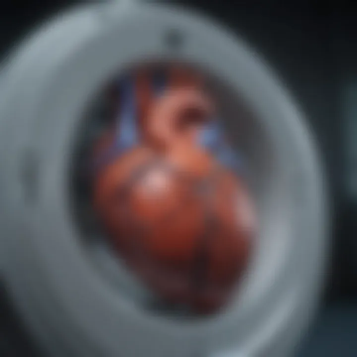
Cardiac imaging has evolved significantly, especially with the introduction of novel techniques such as positron emission tomography-computed tomography (PET CT). This innovative modality combines the functional imaging capabilities of PET with the anatomical detail provided by CT, creating a powerful tool for evaluating cardiovascular health. In recent years, advancements in cardiac PET CT have enhanced its clinical implications, allowing for more precise diagnosis and management of heart disease. By delving into the underlying principles, technological advancements, and specific applications of cardiac PET CT, we can appreciate its role in shaping contemporary cardiac care. This overview aims to illuminate the myriad ways in which cardiac PET CT contributes to the evaluation and treatment of various cardiovascular conditions.
Research Overview
Summary of Key Findings
The research into cardiac PET CT has uncovered several critical insights:
- Enhanced Imaging Resolution: Advances in detector technology have improved spatial resolution, allowing for better visualization of cardiac structures.
- Molecular Imaging Capabilities: PET CT provides unique insights into cardiac metabolism and perfusion, offering invaluable information for diagnosing conditions like coronary artery disease.
- Assessment of Myocardial Viability: Utilizing fluorodeoxyglucose (FDG) in PET CT allows clinicians to better evaluate myocardial viability in patients with heart failure.
Importance of the Research
Understanding cardiac PET CT is vital for multiple reasons. First, it offers a non-invasive approach to visualize heart function and anatomy, significantly reducing patient risk compared to traditional invasive methods. Furthermore, the integration of PET and CT enhances diagnostic accuracy, which can lead to timely and effective treatment interventions. This is particularly important in a landscape where cardiovascular diseases remain a leading cause of morbidity and mortality.
Methodology
Study Design
Research regarding cardiac PET CT typically employs a combination of observational studies, clinical trials, and case series. These designs allow researchers to gather comprehensive data on the efficacy and safety of cardiac PET CT in various clinical scenarios.
Data Collection Techniques
Data is collected through a variety of means:
- Patient Registries: Collecting data from patients undergoing cardiac PET CT for different indications helps build a basis of evidence for its effectiveness.
- Imaging Analysis: Advanced imaging software is used to analyze the data from PET and CT scans, ensuring accurate measurements of perfusion and metabolism.
- Clinical Outcomes Tracking: Follow-up studies assess the long-term impact of findings from cardiac PET CT on patient outcomes, shedding light on its role in management strategies.
Through these methodologies, researchers can validate the clinical relevance of cardiac PET CT in diagnosing and managing heart disease effectively.
As we continue this exploration, we will delve deeper into the specific applications and technical advancements of cardiac PET CT, ultimately revealing its profound implications in the realm of cardiovascular health.
Intro to Cardiac PET CT
Cardiac positron emission tomography-computed tomography, or PET CT, stands as a hallmark innovation in the domain of cardiovascular imaging. This advancement is not just a technical upgrade; it significantly refines diagnostic accuracy and therapeutic strategies in cardiac care. By integrating metabolic and anatomical data, cardiac PET CT yields valuable insights into cardiac function and pathology, which is crucial for effective patient management. With the growing prevalence of heart disease, understanding cardiac PET CT is increasingly important. It allows clinicians to dissect complex cardiac conditions with higher precision.
Historical Context
The evolution of cardiac PET CT can be traced back to the development of positron emission tomography in the late 20th century. Initially limited to oncology and brain imaging, this technology was gradually adapted for cardiology. The first studies applying PET for cardiac purposes surfaced in the 1990s. These early investigations showcased the potential of PET to evaluate myocardial perfusion and viability. At this stage, limited access to the necessary radiotracers hindered widespread adoption.
However, advancements in radiotracer development, coupled with improved CT integration, propelled cardiac PET CT into the forefront of cardiovascular imaging. In the early 2000s, the incorporation of multi-slice CT significantly enhanced anatomical visualization alongside metabolic assessments. This progress marked a shift in how cardiovascular diseases could be diagnosed and treated.
What is Cardiac PET CT?
Cardiac PET CT combines two powerful imaging modalities: positron emission tomography and computed tomography. PET imaging focuses on the metabolic processes within the heart, revealing how well blood flows to different heart regions. CT, on the other hand, provides high-resolution images of the cardiac structures. Together, these technologies allow for a multi-dimensional understanding of cardiac health.
This imaging technique is especially beneficial for diagnosing and assessing coronary artery diseases and other cardiovascular conditions. The ability to visualize blood flow and evaluate the functional state of the myocardium provides critical information for treatment decisions. Moreover, cardiac PET CT contributes to non-invasive management strategies, making it a preferred choice among healthcare providers.
Cardiac PET CT plays a pivotal role in modern cardiovascular diagnostics, bridging anatomical and metabolic assessments, thus enhancing the understanding of various heart conditions.
Through its non-invasive nature and integrated functionality, cardiac PET CT stands as an invaluable tool in contemporary cardiology. As the field continues to evolve, it is vital for practitioners and researchers to stay informed about its capabilities and implications.
Technical Fundamentals of Cardiac PET CT
Understanding the technical fundamentals of cardiac positron emission tomography-computed tomography (PET CT) is essential for professionals working in cardiovascular health. This section addresses the mechanisms that drive PET imaging and its integration with CT imaging. By understanding these foundational concepts, practitioners can better utilize cardiac PET CT in clinical practice.
The Mechanism of PET Imaging
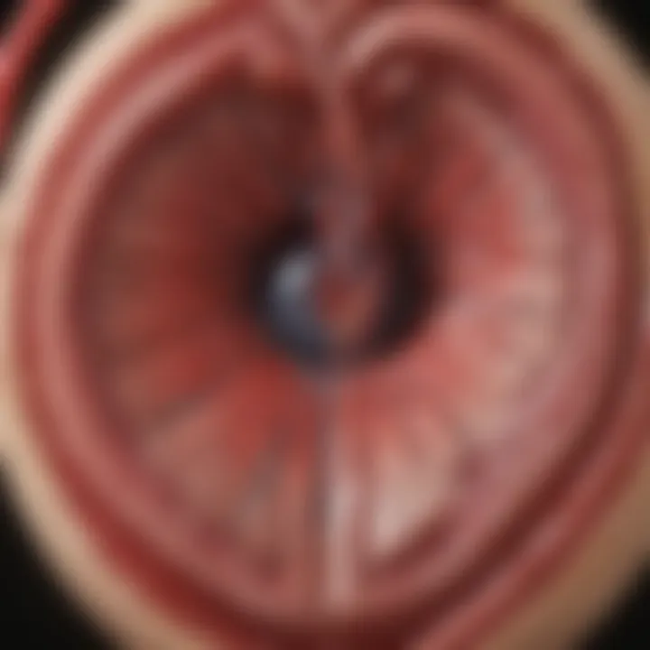
Positron emission tomography is a nuclear imaging technique. It creates images based on functional processes within the body, specifically, the metabolic activities of tissues. In cardiac PET imaging, radiotracers are injected into the patient. Once in the bloodstream, these tracers emit positrons as they decay.
When a positron encounters an electron in the body, they annihilate each other, producing two gamma photons that travel in opposite directions. These photons are detected by the PET scanner, which calculates their original location based on the timing of their detection. Modern PET systems employ sophisticated algorithms to reconstruct detailed images of the heart, clearly illustrating metabolic activity.
This imaging technique is particularly useful in assessing cardiac conditions as it evaluates myocardial perfusion and metabolism. The most commonly used radiotracers in cardiac PET include fluorodeoxyglucose (FDG) for assessing glucose metabolism and ammonia for evaluating blood flow.
Integration with CT Imaging
The integration of PET with computed tomography enhances the capabilities of cardiac imaging. CT imaging provides high-resolution anatomical data, which complements the functional information gathered by PET. This fusion results in hybrid imaging, allowing for a more comprehensive understanding of cardiac conditions.
In cardiac PET CT, the sequences are often acquired simultaneously. The patient lies still while the PET and CT scans are performed. The CT scan captures detailed structural images of the heart, providing context for the functional images obtained through PET. This combination helps differentiate between viable and non-viable myocardium, aiding in decision-making processes regarding treatment options.
The dual capabilities of PET CT influence clinical practice in significant ways. It allows for accurate localization of lesions and interpretation of perfusion abnormalities more precisely, improving diagnosis and management strategies for conditions like coronary artery disease.
"The integration of PET and CT has revolutionized cardiac imaging, providing unparalleled insight into both function and structure."
In summary, comprehending the mechanisms of PET imaging and the advantages of its integration with CT imaging is crucial. These technical fundamentals form the backbone of cardiac PET CT and support its growing relevance in cardiovascular diagnostics.
Clinical Applications of Cardiac PET CT
The integration of cardiac positron emission tomography-computed tomography (PET CT) within cardiovascular medicine has revealed a multitude of clinical applications. This imaging technique provides essential insights into the pathophysiology of heart diseases, enabling tailored treatment approaches. The ability of cardiac PET CT to evaluate metabolic and perfusion characteristics of the myocardium facilitates precise diagnoses and prognosis in various cardiac conditions. As such, comprehending these applications is vital for professionals aiming to optimize patient care.
Diagnosis of Coronary Artery Disease
Coronary artery disease (CAD) remains a leading cause of morbidity and mortality globally. Cardiac PET CT plays a crucial role in diagnosing CAD through its ability to assess myocardial perfusion. This imaging modality gauges blood flow in the pericardial arteries, revealing regions of ischemia that might not be evident in traditional imaging methods. By utilizing radiotracers such as fluorodeoxyglucose (FDG), clinicians can obtain detailed images that reflect myocardial metabolism. This metabolic assessment allows differentiation between viable and non-viable myocardial tissue.
One notable benefit of cardiac PET CT is its superior sensitivity in detecting coronary artery lesions, especially in cases where non-invasive tests like stress tests yield inconclusive results. The combination of both functional and anatomical data enables a comprehensive evaluation, thereby enhancing diagnostic accuracy.
Assessment of Myocardial Viability
Understanding myocardial viability is essential when planning interventions for patients with ischemic heart disease. Cardiac PET CT aids in identifying viable myocardium that can potentially recover following revascularization. In clinical practice, the amount of viable myocardium determines the therapeutic strategy, influencing decisions about coronary artery bypass grafting or percutaneous coronary intervention.
The assessment typically involves imaging the heart after administering FDG, a glucose analog that accumulates in metabolically active tissues. When regions of the heart exhibit reduced FDG uptake, it signifies the presence of necrotic tissue, which is generally non-functional. Conversely, areas showing substantial uptake suggest viable tissue, highlighting the potential benefit of revascularization. This vital information directly influences treatment strategies and patient prognosis, making PET imaging an indispensable tool in managing myocardial viability.
Evaluation of Heart Failure
Heart failure is a complex syndrome with numerous underlying causes. The role of cardiac PET CT in this context focuses on determining myocardial perfusion and metabolism, crucial in understanding the disease's etiology. By evaluating regional blood flow and myocardial function, practitioners can gauge the severity and potential reversibility of heart failure.
In patients with heart failure, cardiac PET CT assists in differentiating between various types of cardiac dysfunction, such as ischemic versus non-ischemic causes. This distinction is fundamental, as it influences both management decisions and prognosis.
Furthermore, the ability to assess myocardial perfusion during stress testing enhances the evaluation of cardiac function, allowing clinicians to determine the most appropriate treatment strategy, whether it's medical management, device therapy, or surgical intervention.
Cardiac PET CT's non-invasive nature and its insightful diagnostic capabilities make it a leading choice in the cardiovascular landscape.
In summary, the clinical applications of cardiac PET CT are paramount in heart disease management. From diagnosing CAD and assessing myocardial viability to evaluating heart failure, this advanced imaging technique serves as a cornerstone for informed decision-making in cardiovascular care.
Molecular Imaging and Cardiac PET CT
Molecular imaging plays a crucial role in enhancing the understanding of cardiac conditions and the overall functionality of the heart. Cardiac PET CT merges the strengths of metabolic imaging with anatomical insights, offering a comprehensive approach to diagnosing and managing heart diseases. This integration of technologies allows clinicians to view both the structure and the function of the heart concurrently, ultimately leading to better-informed treatment decisions.
Advancements in molecular imaging techniques facilitate not only the evaluation of coronary artery disease and myocardial viability but also provide insights into the underlying pathways of cardiac diseases. The capability to visualize metabolic processes in the heart underscores the importance of Cardiac PET CT in contemporary medical practice. As diseases progress, their metabolic profiles change; hence by understanding these changes, healthcare professionals can tailor interventions accordingly.
"Molecular imaging techniques allow us to directly visualize and quantify biological processes in tissues, leading to earlier and more accurate diagnosis of cardiac diseases."
Understanding Metabolic Imaging
Metabolic imaging focuses on assessing the biochemical processes within the heart tissue. In the context of Cardiac PET CT, it utilizes specific radiotracers that highlight the metabolism of myocardial cells. By measuring the uptake of these radiotracers, clinicians can determine how well the heart tissue is functioning and whether it is receiving adequate blood supply.
For example, fluorodeoxyglucose (FDG) is one of the primary radiotracers used in metabolic imaging. FDG uptake correlates with glucose metabolism, which changes in various cardiac conditions such as ischemia or heart failure. By quantifying the metabolic activity of the myocardial tissue, medical professionals can make informed decisions regarding further diagnostic steps or treatments.
Key Radiotracers in Cardiac PET
Several key radiotracers are fundamental to the functionality of cardiac PET imaging. Each of these agents provides vital information regarding different aspects of heart health.
- Fluorodeoxyglucose (FDG): Used for assessing glucose metabolism, especially useful in evaluating myocardial viability and detecting inflammation in coronary artery disease.
- Ammonia-13: This radiotracer is beneficial in assessing myocardial perfusion. It helps depict blood flow through the heart muscle during rest and stress conditions.
- Rubidium-82: Another agent used for measuring myocardial perfusion, known for its high resolution and rapid clearance from the body, which aids in capturing dynamic changes in blood flow.
In summary, the integration of these radiotracers within Cardiac PET CT not only enhances diagnostic capabilities but also enriches the understanding of the etiology and progression of heart diseases. This detailed molecular perspective is increasingly vital for developing personalized treatment strategies.
Comparative Analysis with Other Imaging Modalities
The comparative analysis of cardiac PET CT with other imaging modalities is vital for understanding its specific advantages and applications. It allows professionals to discern which techniques may best meet the diagnostic needs of individual patients. Given the complexity of cardiovascular diseases, selecting the appropriate imaging modality can greatly influence treatment outcomes. The two primary modalities for comparison are single photon emission computed tomography (SPECT) and magnetic resonance imaging (MRI). Each of these approaches has its unique strengths and limitations, which must be carefully considered to optimize patient care.
Cardiac PET CT vs. SPECT
Cardiac PET CT and SPECT both use nuclear imaging to assess cardiac conditions. However, they differ significantly in their technology and applications. Cardiac PET CT offers superior spatial resolution and sensitivity. It provides clearer and more accurate images of cardiac blood flow and metabolism when compared to SPECT. This is primarily due to the higher energy photons emitted by the radiotracers used in PET imaging.
In addition, PET CT can simultaneously provide functional and anatomical information. It integrates the metabolic data from PET with the detailed anatomical structure from CT scans. This dual capability is advantageous in many clinical scenarios.
Key Differences:
- Sensitivity and Resolution: Cardiac PET CT exhibits higher sensitivity and resolution than SPECT, which can lead to more reliable diagnoses.
- Radiotracers: PET typically uses radiotracers such as fluorodeoxyglucose (FDG), while SPECT often uses technetium-99m based tracers.
- Imaging Time: Cardiac PET scans typically require less time than those conducted with SPECT, improving patient comfort.
Despite these advantages, SPECT imaging is often more accessible and less expensive, making it a practical choice in various clinical settings. Therefore, the choice between cardiac PET CT and SPECT largely hinges on factors such as availability, cost, and clinical indication.
Cardiac PET CT vs. MRI
When comparing cardiac PET CT to MRI, it is essential to note the differences in how each modality visualizes the heart. MRI employs powerful magnets and radio waves to generate images, providing exceptional anatomical detail and tissue characterization. However, MRI is less effective in assessing myocardial perfusion when compared to PET.
PET CT's advantage lies in its ability to assess metabolic information directly, enabling clinicians to evaluate not only the structure but also the essential functions of the heart.
Considerations for Selection:
- Anatomical Detail: MRI provides superior anatomical resolution, particularly in evaluating complex structural heart diseases.
- Functional Assessment: On the other hand, PET CT excels in functional studies, particularly in assessing myocardial perfusion and viability.
- Patient Compatibility: MRI may be contraindicated in patients with implanted devices or metal allergies, limiting its use in certain populations. Conversely, PET CT is safe in such cases.
"Understanding the nuances between these modalities is essential for clinicians to deliver optimal patient-centered care."
Benefits of Cardiac PET CT
Cardiac positron emission tomography-computed tomography (PET CT) offers numerous benefits in the diagnostic and management landscape of cardiovascular diseases. This section elaborates on the importance of these advantages, specifically highlighting the non-invasive nature of the technique as well as the valuable insights it provides into disease mechanisms. Recognizing these benefits enhances the understanding of cardiac PET CT's role and its impact on patient care.
Non-Invasiveness
One of the most significant advantages of cardiac PET CT is its non-invasive nature. Unlike traditional cardiac procedures that may require catheterization or surgical intervention, cardiac PET CT allows for comprehensive imaging without the need for any invasive measures. This is particularly beneficial for patients at high risk or those with comorbidities that make invasive procedures dangerous.
Furthermore, the non-invasive aspect encourages patient cooperation and comfort. Patients may experience less anxiety when they know that the procedure does not involve needles or surgery. This comfort can lead to more accurate diagnostic results, as the patients are likely to adhere better to the imaging protocols when they do not feel stress from invasive techniques.
Insights into Disease Mechanisms
Cardiac PET CT provides critical insights into the underlying mechanisms of various cardiovascular diseases. By utilizing innovative radiotracers, this imaging technique allows for the evaluation of cardiac metabolism and perfusion. Understanding these factors is crucial for developing effective treatment plans tailored to the specific needs of each patient.
The ability to visualize metabolic processes offers cardiologists a deeper understanding of conditions like ischemic heart disease, heart failure, and myocardial viability. For instance, analyzing glucose metabolism can help differentiate between viable heart tissue and areas of scar. This differentiation directly influences treatment decisions, such as whether to proceed with surgical interventions or medical management. Additionally, with the integration of PET CT data into clinical workflows, professionals can monitor disease progression and treatment responses over time.
“Cardiac PET CT allows for non-invasive exploration of metabolic processes, providing insights that are pivotal in personalized patient care.”
Overall, the benefits of cardiac PET CT significantly advance the field of cardiovascular health, offering a less invasive method of diagnosis while enhancing the understanding of complex disease mechanisms. These advantages underscore the relevance of cardiac PET CT in modern clinical practice and highlight its integral role in the future of cardiac care.
Limitations and Challenges
Understanding the limitations and challenges of cardiac positron emission tomography-computed tomography (PET CT) is vital in evaluating its overall efficacy in clinical settings. While this technology offers numerous benefits in terms of non-invasive imaging, it faces significant hurdles that can impact its application in routine practice. This section aims to elucidate both cost implications and technical limitations associated with cardiac PET CT. Recognizing these factors is essential for any professional in the field, as they directly influence both patient care and the decision-making process in cardiology.
Cost Implications
One of the primary concerns surrounding cardiac PET CT is its cost. The initial setup for such imaging technology can be quite substantial. Facilities must invest in specialized equipment, which requires ongoing maintenance and calibration. In addition, the high cost of radiotracers that are used in PET imaging adds another layer of expense. This can create barriers for many healthcare facilities, particularly those in less developed regions or with limited resources.
Patients may also face high out-of-pocket expenses, which can discourage them from choosing this imaging modality. Reimbursement rates from insurance providers for procedures involving cardiac PET CT are often less favorable compared to those for other imaging techniques, such as echocardiography or even cardiac MRI. This creates a financial disincentive for clinicians who may otherwise consider recommending PET CT for their patients.
In summary, the significant costs associated with cardiac PET CT can hinder its accessibility and uptake, raising questions about its practicality in everyday clinical practice.
Technical Limitations
Technical limitations of cardiac PET CT encompass various aspects of the imaging process that can affect the quality of results. One notable issue is the temporal resolution of PET imaging. While PET CT can provide functional information about the heart, its imaging speed may not always align with fast physiological processes, making it less effective for capturing transient changes in cardiac function compared to other modalities.
Another technical consideration is the limitations in spatial resolution. Although advancements have been made, the detail obtainable in PET imaging may not be as fine as what some fully dedicated cardiac imaging modalities offer. This can influence the accuracy of diagnosing smaller pathological changes in cardiac tissue, potentially compromising the quality of patient assessment.
Furthermore, variability in patient body habitus can lead to inconsistencies in image quality. Obesity or irregular heart rates can produce artifacts that may mislead interpretation or complicate diagnosis.
Future Directions in Cardiac PET CT
The realm of cardiac positron emission tomography-computed tomography (PET CT) is on the brink of remarkable advancements. This section outlines the future directions that not only hold promise but also challenge the current paradigms in cardiovascular imaging. Emphasis on innovation and the evolution of clinical guidelines are integral to understanding how these technologies can further enhance patient outcomes and clinical efficiency.
Innovative Technologies on the Horizon
New technologies are continuously emerging to improve the capabilities of cardiac PET CT. One notable direction is the integration of artificial intelligence (AI) in image analysis. AI can automate image interpretation, reducing the burden on radiologists and improving diagnostic accuracy. Enhanced algorithms will assist in identifying subtle abnormalities that may be overlooked through traditional methods.
Another key advancement is the development of novel radiotracers that can provide more specific information about cardiac function and metabolism. For example, tracers that target specific biological markers associated with heart disease are being researched. These have potential to provide insights beyond what is currently possible with standard imaging protocols.
Emerging hybrid imaging systems also play a significant role in future developments. Devices that combine PET with other imaging modalities such as MRI can lead to a more comprehensive understanding of cardiac conditions. This hybrid technology may help in visualizing both anatomical and functional details, offering a more complete picture for clinicians.
Enhancing Clinical Guidelines
As advancements in cardiac PET CT continue, it is crucial to refine clinical guidelines surrounding its use. With new technologies and methodologies, protocols must adapt to ensure they meet the evolving landscape of patient care. Incorporating findings from ongoing research into guideline updates will help clinicians apply the most effective imaging techniques in practice.
Collaboration between cardiologists, radiologists, and researchers is vital for establishing robust evidence-based guidelines. This includes creating standards for new imaging protocols and specific indications for the use of cardiac PET CT in various patient populations.
Continuous education for healthcare professionals on these advancements is another important factor. By keeping practitioners informed, it can help transition new technologies from research settings to routine clinical use. This facilitates the efficient application of cutting-edge imaging strategies in everyday practice, ultimately enhancing patient management.
"The collective advancement in medical imaging relies on the synergy of technology, clinical application, and ongoing education in the healthcare community."
In summary, future directions of cardiac PET CT are filled with potential. Innovations in technology and the refinement of clinical guidelines will shape how this imaging modality impacts cardiovascular care. Embracing these changes will be essential for optimizing patient outcomes and advancing clinical practices.
Finale
In the rapidly evolving landscape of cardiac imaging, the conclusion of this article underscores the significant role that cardiac positron emission tomography-computed tomography (PET CT) plays in enhancing cardiovascular care. This imaging method offers unique capabilities that benefit both diagnosis and therapeutic decision-making. Understanding these implications is essential not only for practitioners in the field but also for researchers advancing cardiac imaging technologies and methodologies.
Summarizing Key Insights
Throughout this article, we have dissected various aspects of cardiac PET CT. Here are some key points to remember:
- Versatility in Applications: Cardiac PET CT is instrumental in diagnosing coronary artery disease, assessing myocardial viability, and evaluating heart failure. Its capability to visualize both physiological and metabolic functions of the heart is a strong asset.
- Enhanced Imaging Quality: The integration of PET with CT provides high-resolution images and quantitative analysis. This combination delivers comprehensive insights into cardiac health that other modalities might overlook.
- Molecular Imaging Advantage: The use of specific radiotracers allows for detailed analysis of heart metabolism and perfusion. This level of detail aids in formulating precise treatment strategies tailored to individual patient needs.
- Future Directions: As technology progresses, future advancements promise to enhance the accuracy, efficiency, and cost-effectiveness of cardiac PET CT, expanding its accessibility and utility in clinical settings.
Final Thoughts on Implementation
Implementing cardiac PET CT into clinical practice requires thoughtful consideration of several factors. It is vital to ensure that practitioners are well-trained in interpreting PET CT results and understanding their implications for patient management.
Moreover, institutions must consider the cost implications outlined previously. Initiatives aimed at reducing barriers related to cost and accessibility are essential to foster wider adoption of this advanced imaging technology.
Finally, ongoing education regarding the latest advancements in cardiac PET CT will enable healthcare professionals to stay informed about best practices and emerging opportunities in patient care. Proper integration of this imaging technology into clinical workflows can potentially lead to improved outcomes for patients with cardiovascular diseases.
Ultimately, the continued growth and refinement of cardiac PET CT will likely play a pivotal role in shaping the future landscape of cardiovascular healthcare.
"Cardiac PET CT not only enhances diagnostic accuracy but also opens avenues for personalized treatment plans, representing a vital stride in modern cardiovascular medicine."
For further resources on cardiac imaging advancements, visit Wikipedia or Britannica.
Keep an eye on community discussions about this topic on platforms like Reddit and Facebook to gain insights from peers.


