Cochlear Bones: Anatomy, Function, and Health Impact
Intro
Cochlear bones, though often overlooked, are fundamental to our ability to hear. They form part of a complex system that transmits sound waves, translating vibrations into signals that our brain decodes as sound. Their intricate anatomy is fascinating, comprising several small and delicate structures that work together seamlessly. Understanding these components is essential not just for those in the field of audiology but also for anyone interested in how we experience the world through sound.
Research Overview
Summary of Key Findings
Recent studies have shed light on several crucial aspects of cochlear bones. Researchers have identified that these bones consist mainly of the cochlea, the stapes, incus, and malleus. The cochlea itself is coiled and filled with fluid, while the stapes acts as a bridge to the inner ear, connecting sound waves to sensory receptors. The delicate balance in their function is vital; any deformation or damage can significantly impair hearing.
Importance of the Research
The significance of exploring cochlear bones goes beyond academic interest. Many individuals suffer from hearing loss due to various conditions, including otosclerosis, ossicular chain discontinuity, or congenital malformations. By understanding the anatomy and functions of these bones, medical professionals can improve diagnosis and treatment plans, making life easier for those affected. As technology advances, surgical techniques also evolve, offering new hope for restoration of hearing.
Methodology
Study Design
The research into cochlear bones typically employs a combination of observational and experimental approaches. Studies often leverage advanced imaging techniques to visualize the cochlear structures in living subjects, combined with cadaver examinations for a more detailed anatomical understanding.
Data Collection Techniques
Data on cochlear function and health may be gathered through various methods, including audiometric tests, imaging studies like MRI or CT scans, and surgical observations during procedures aimed at correcting issues related to cochlear bones. These methods together allow researchers to build a comprehensive picture of how the cochlear bones operate within the auditory system.
The health of cochlear bones is crucial in maintaining normal hearing. Their intricate anatomy supports the complex process of sound transmission, making them a focal point in audiology research.
As we delve deeper into the complexities of cochlear bones, we open the door to understanding not only their structure but also the profound implications for auditory health and the future of treatments for hearing loss.
Foreword to Cochlear Bones
Cochlear bones form an integral part of the auditory system, serving as the foundational structures within the cochlea, a spiraled organ responsible for transforming sound vibrations into neural signals. Understanding cochlear bones is crucial for several reasons. For one, they are the unsung heroes that facilitate the delicate hearing process. Trouble or changes in these bones can lead to significant hearing impairments, impacting day-to-day communication and overall quality of life.
Overview of the Cochlea
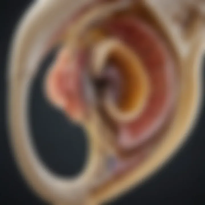
The cochlea resembles a snail shell in its structure, curved and compact with several compartments, among which the osseous labyrinth is prominent. Within this labyrinth reside the cochlear bones, which include the modiolus, scala vestibuli, and scala tympani. While these bones may appear to the untrained eye as mere structural components, they actually possess a complex design that efficiently supports the mechanics of hearing.
The cochlea houses hair cells, sensory receptors that convert sound waves into electrical impulses. As sound waves travel through the air, they enter the outer ear and eventually cause vibrations in the cochlear bones. This facilitates their movement and plays a pivotal role in sound transmission. The cochlea's architecture is a testimony to evolution's ingenuity, demonstrating how biological systems have adapted over time to foster effective communication through auditory pathways.
Importance of Cochlear Bones in Hearing
Understanding the role of cochlear bones is akin to comprehending the gears in a finely-tuned clock. Their health is not merely beneficial; it's essential. Here's why:
- Sound Manipulation: Cochlear bones provide the necessary environment for sound waves to be transformed into signals the brain can interpret. If there's even a slight misalignment or degeneration, it can result in auditory deficits that may not only affect hearing but also emotional and mental well-being.
- Supportive Framework: These bones also serve as structural supporters for sensory cells within the cochlear canals. This relationship is symbiotic; while the bones provide a stable platform for sensory cells to function, the sensory cells in turn relay critical auditory information back to the brain.
The relevance of cochlear bones extends well beyond mere anatomy. They are essential in several medical discussions regarding hearing loss and treatment avenues, like cochlear implants or surgical interventions. For those studying auditory systems, the pursuit to understand these bones can lead to new insights and methods of both diagnosis and treatment of hearing loss, ensuring that communication remains a fundamental human experience.
The cochlear bones may be small in size, but their importance in hearing functionality is absolutely monumental.
Anatomy of Cochlear Bones
Understanding the anatomy of cochlear bones is paramount in appreciating their role in hearing. These structures, nestled within the cochlea, contribute not only to the transmission of sound but also to overall auditory health. By dissecting the various components of these bones, we can unveil how they interact with other elements of the auditory system and why their integrity is essential for effective hearing.
Structure of the Cochlear Osseous Labyrinth
The cochlear osseous labyrinth is a bony structure resembling a snail shell, coiling around a central axis known as the modiolus. Its unique spiral shape allows for an intricate arrangement of auditory components. This spiral structure protects delicate sensory cells while providing a passage for sound waves to travel through the cochlea.
A noteworthy aspect of the osseous labyrinth is its division into three main parts: the scala vestibuli, scala tympani, and the cochlear duct. These sections play distinct roles in transforming sound vibrations into neural signals. The bony walls of the cochlea house the organ of Corti, where sensory hair cells detect the sound waves, converting them into signals sent to the brain for processing.
In summary, the coherence of the structure is integral for the efficient functioning of the auditory system. Any compromise in the structural integrity may lead to disorders affecting hearing.
Types of Cochlear Bones
The cochlea comprises several types of bones, each contributing valuable functions that are interlinked in the process of hearing.
Scala vestibuli
The scala vestibuli, located at the upper part of the cochlea, is crucial for conducting sound waves from the oval window to the cochlear duct. This chamber is filled with perilymph, a fluid that helps transmit sound vibrations effectively. One of the defining characteristics of the scala vestibuli is its stiffness, which enhances the ability to transmit high-frequency sounds.
The unique feature of the scala vestibuli lies in its connection to the vestibular system, which maintains balance and spatial orientation. This juxtaposition makes it vital not just for hearing but for overall spatial awareness. However, if this bone is compromised, issues like vertigo and imbalance may ensue, illustrating why maintaining its health is essential.
Scala tympani
Positioned below the cochlear duct, the scala tympani mirrors the scala vestibuli’s anatomical structure and function. It too is filled with perilymph, providing a crucial environment for sound wave transmission. One remarkable characteristic of the scala tympani is its role in dissipating sound wave energy through the round window.
What sets the scala tympani apart is its function in aiding the cochlear fluid dynamics during sound transmission. It allows for the proper pressure release and prevents damage to the cochlear structures. Understanding its function helps explain why any obstruction or damage here can lead to conductive hearing loss, emphasizing the importance of this component in auditory health.
Modiolus
The modiolus serves as the central pillar of the cochlea, entwining with the scalae to provide structural support. Additionally, it houses the spiral ganglia, where sensory neurons reside, transmitting impulses from the cochlea to the brain. Its importance cannot be overstated; without a healthy modiolus, the transmission of sound information would be significantly compromised.
A defining characteristic of the modiolus is its rich vascular supply, enabling it to maintain the health of surrounding tissues. This unique feature highlights the modiolus as a hub of neural connectivity, tying together auditory pathways. Likewise, any degeneration here can lead to profound sensory neural hearing loss, stressing the importance of its preservation in hearing health.
Functions of Cochlear Bones
Cochlear bones serve several critical roles in the auditory system, often overlooked but exceedingly important for effective hearing. Their functions encompass sound wave transmission and support for sensory cells, making them essential for our ability to perceive sound accurately. Understanding these functions can help appreciate how integral cochlear bones are in the broader auditory landscape.
Sound Wave Transmission
Sound waves are vibrations that travel through the air, entering the ear canal before they meet the tympanic membrane, commonly known as the eardrum. Once the eardrum vibrates in response to these sound waves, it sets off a series of mechanical actions involving the ossicles, small bones located within the middle ear, which connect the eardrum to the cochlea. However, it is primarily in the cochlea where the osseous labyrinth, consisting of cochlear bones like the scala vestibuli and scala tympani, plays an irreplaceable role.
Here’s a breakdown of how cochlear bones aid in sound wave transmission:
- Vibration Amplification: The cochlear bones help amplify the sound vibrations initiated by the ossicles. This amplification is critical because it allows quieter sounds to be discernible, effectively increasing sensitivity to auditory input.
- Fluid Movement: Sound waves, after being amplified, move through the fluid-filled chambers of the cochlea. Cochlear bones facilitate this fluid movement, which is vital for translating mechanical vibrations into electrical signals that the auditory nerve can interpret.
- Frequency Separations: Different regions of the cochlea respond to different sound frequencies due to the unique structure of cochlear bones. This separation is fundamental for sound differentiation; high-frequency sounds affect certain areas of the cochlear base while lower frequencies resonate deeper within the cochlea.
"Understanding the mechanics of sound wave transmission is crucial for diagnosing and treating hearing impairments."
Support for Sensory Cells
Another pivotal function of cochlear bones is providing a structural framework and support for sensory cells, particularly the hair cells located in the organ of Corti. These specialized cells are the sensory receptors for sound, and their function relies heavily on the integrity and stability offered by the cochlear bones.
The support offered by cochlear bones materializes in various ways:
- Physical Stability: The rigid structure of the cochlear bones maintains a stable environment for the delicate hair cells, preventing displacement during the energetic process of sound transmission. If these bones weaken or become damaged, the hair cells may lose their integrity, hampering their ability to detect sound waves.
- Pressure Distribution: During sound wave transmission, cochlear bones assist in evenly distributing pressure throughout the cochlear fluid. This distribution is essential for the proper functioning of sensory cells, enabling them to respond accurately to sound stimuli.
- Microbial Defense: Healthy cochlear bones can also play a part in defending against infections, particularly those that might impede the function of sensory cells. The structural integrity of these bones prevents the intrusion of pathogens, ensuring auditory health.
Health Implications of Cochlear Bones
Understanding the health implications of cochlear bones is essential in grasping their role in hearing and overall auditory health. Cochlear bones, while often overlooked, are fundamental in maintaining the structure and function of the auditory system. The implications of their health extend beyond simple anatomy; they touch upon significant medical conditions, treatment options, and the advancements in auditory health care. As the framework that supports our hearing mechanism, any changes or afflictions to cochlear bones can lead to profound consequences for auditory capabilities.
Impact of Otosclerosis
Otosclerosis is a condition that can significantly affect cochlear bones, primarily characterized by abnormal bone growth in the middle ear. This condition primarily targets the stapes bone, but it also has implications for the cochlea itself. When the stapes becomes anchored in place due to this abnormal growth, sound waves encounter difficulties reaching the inner ear. As a result, conductive hearing loss often ensues, meaning that sounds cannot be adequately transmitted through the auditory pathway.
Some of the symptoms include:
- Hearing loss
- Tinnitus, a persistent ringing in the ears
- Balance issues due to compromised auditory function
In more severe cases, otosclerosis can disrupt the delicate interactions within both the scala vestibuli and scala tympani, altering fluid dynamics in the cochlea. By interfering with the normal movement of perilymph, the effects ripple back through the auditory system, leading to further hearing impairments. This underscores the importance of early detection and intervention to prevent long-term damage.
Effects of Age-Related Changes
As individuals age, a host of biological changes occur, and cochlear bones are not immune to this natural process. Age-related changes can lead to the thinning and loss of density in the bony structures of the cochlea. Such changes alter the ability of cochlear bones to effectively transmit sound waves, causing both conductive and sensory neural hearing loss.
In particular, studies have noted that:
- The overall integrity of the cochlear osseous labyrinth diminishes over time
- The modiolus, which serves as a central core for nerve fibers, may also become less robust, affecting auditory signal transmission
- Fluid movement within the cochlear cavities becomes less efficient, further complicating sound processing
These age-related changes highlight the need for ongoing research into protective measures and treatment options for the aging auditory system. Understanding the intersection of cochlear health and age is crucial for developing improved interventions and maintaining auditory health over an individual's lifetime.
"Aging is not lost youth, but a new stage of opportunity and strength."
— Betty Friedan
Cochlear Bones and Hearing Loss
Cochlear bones stand as a crucial bridge between understanding the sounds of the world and the mechanisms that can lead to hearing loss. They provide both structural and functional support, essentially functioning as the foundation where hearing begins. If something disrupts this intricate system, the results can be profound, often leading to various forms of hearing impairment. Grasping the relationship between cochlear bones and hearing loss not only sheds light on the severity of auditory issues but also paves the way for effective treatments and interventions.
Types of Hearing Loss Related to Cochlear Bones
Hearing loss stemming from cochlear bones generally falls into two categories: conductive and sensory neural. Each type encompasses specific characteristics and implications, allowing for a more complete understanding of auditory health.
Conductive Hearing Loss
Conductive hearing loss arises when sound waves are obstructed from reaching the inner ear. This type of loss is primarily linked to issues with the outer or middle ear, including problems with the cochlear bones. A key characteristic of conductive hearing loss is the reduction in the sound pressure level reaching the cochlea.
The benefits of focusing on conductive hearing loss in this article are substantial:
- Identification of Obstructions: Understanding this type helps highlight how conditions like otosclerosis can affect bone structures, leading to defined auditory issues.
- Treatment Opportunities: Since conductive hearing loss often responds well to medical or surgical interventions, understanding its connection with cochlear bones opens doors for patients seeking relief.
A unique feature of conductive hearing loss is that it can often be treated effectively, allowing for the restoration of hearing through procedures like stapedectomy. However, it may also vary in severity, leading to a spectrum of auditory challenges.
Sensory Neural Hearing Loss
In contrast, sensory neural hearing loss is often characterized by damage to the inner ear structures and subsequently the auditory nerve. This kind of loss is tricky because it involves the sensory cells and auditory pathways that process sounds, often linked to the cochlear function. One significant aspect of sensory neural hearing loss is that it can result from aging, trauma, or exposure to loud noises.
Discussing sensory neural hearing loss is equally relevant as it:
- Emphasizes Long-Term Damage: This type of hearing loss can be irreversible due to nerve damage, marking it as a serious concern for auditory health.
- Awareness of Cochlear Bones’ Role: It illustrates how important cochlear anatomy is for sound perception, guiding research and clinical approaches towards developing effective interventions.
A unique feature here is the subtlety of its symptoms, often resulting in gradual hearing degradation, which can go unnoticed until significant loss occurs. Unfortunately, the options for restoration are limited, putting more emphasis on prevention and early intervention.
Cochlear Implants: Mechanism and Impact
Cochlear implants serve as a beacon of hope for those grappling with various types of hearing loss, integrating tightly with the conversation around cochlear bones. These devices bypass damaged structures, providing direct stimulation to the auditory nerve, effectively restoring a sense of sound. The mechanism of cochlear implants involves converting sound waves into electrical signals that can be interpreted by the brain.
The impact of cochlear implants is profound:
- Restoration of Hearing: Many individuals who otherwise would face profound hearing loss can regain significant auditory capabilities, markedly improving their quality of life.
- Technological Advances: Ongoing developments in implant technology continue to enhance outcomes, providing tailored solutions based on specific patient needs.
As we continue to explore the many dynamics within cochlear anatomy, it’s clear that cochlear bones are not just a mere structure; they are fundamentally intertwined with the very nature of hearing itself.
Research and Advancements
Research into cochlear bones is pivotal, as it not only uncovers the intricacies of auditory anatomy but also drives innovations in treatment methods. Understanding the cobweb of connections within the cochlea can lead to improved therapeutic strategies against hearing loss. With the ever-advancing field of audiology, current research focuses on several key areas that illuminate the role of cochlear anatomy in auditory health and treatment options.
Current Research Focuses
Scientists are honing in on various aspects of cochlear bones that influence hearing capabilities. Key areas of exploration include:
- Biomaterials for Regeneration: There's ongoing research into utilizing biocompatible materials to aid in the regeneration of damaged cochlear structures. This could potentially restore hearing in patients with severe cochlear damage.
- Mechanisms of Otosclerosis: Investigating how otosclerosis affects the bony labyrinth can unveil new treatment pathways to combat this condition, which is notorious for causing hearing impairment.
- Synergy of Cochlear Structures: Delving into how cochlear bones interact with other auditory components, such as hair cells, is showing promise. Such studies may inform the development of interventions that enhance or mimic these natural interactions.
Research in these directions is crucial not just for understanding the basic science of hearing but for applying this knowledge in clinical settings.
Innovations in Cochlear Implant Technology
The evolution of cochlear implants exemplifies the remarkable progress in auditory technology. Today’s cochlear implants are more sophisticated than ever, leveraging advanced engineering and biological insights to enhance their efficacy.
"Innovations in cochlear implants are increasingly transforming the landscape of auditory restoration, providing renewed hope for those with severe hearing loss."
Some noteworthy advancements include:
- Reduced Size and Enhanced Design: Modern devices are smaller, making surgeries less invasive and recovery quicker. The integration of thin film technologies allows for better performance in a more compact format.
- Improved Sound Processing: New algorithms analyze and process sound more efficiently, enabling clearer audio experiences for users in diverse environments.
- Wireless Technology Integration: Recent implants often connect wirelessly to smartphones or other devices, allowing users to stream audio directly and customize their hearing experience.
These innovations not only bolster the effectiveness of implany technology but also signify a substantial shift toward personalized auditory solutions. As research continues to unearth the complexities of cochlear anatomy, the future of hearing restoration looks promising.
Surgical Techniques and Interventions


Surgical techniques and interventions related to cochlear bones play a crucial role in addressing various auditory disorders. These procedures not only aim to restore hearing but also help mitigate the effects of conditions affecting the cochlear area. Pioneering methods such as mastoidectomy and cochlear implant fittings have become essential components in contemporary otologic practice. The benefits of these interventions range from improved auditory function to enhanced quality of life for patients suffering from hearing loss.
Mastoidectomy and Its Implications
Mastoidectomy involves the surgical removal of the mastoid air cells located behind the ear. This procedure can become necessary in cases where chronic infections or extensive cholesteatoma are present. The importance of mastoidectomy cannot be overstated; it addresses infection, prevents further complications, and aids in better access to the cochlear structures.
- Indications for Mastoidectomy:
- Persistent otitis media (middle ear infections)
- Cholesteatoma, a destructive ear condition
- Complications involving the skull base
By removing infected tissue, surgeons can also improve the overall environment for cochlear structures. This fosters healing and may subsequently enhance the effectiveness of other interventions such as cochlear implants.
"Mastoidectomy can significantly increase the chances of success for patients requiring cochlear implants, thus bridging the gap between conductive and sensorineural hearing losses."
Fitting Cochlear Implants: Steps and Considerations
Fitting cochlear implants is a meticulous process that demands careful planning and execution. This intervention is particularly vital for individuals with severe to profound hearing loss, who may not benefit from traditional hearing aids.
The fitting procedure generally consists of several crucial steps:
- Pre-Operative Assessment:
- Surgical Procedure:
- Post-Operative Rehabilitation:
- Comprehensive auditory evaluation
- Eligibility determination through imaging techniques, like CT scans
- Counseling to set realistic expectations.
- The implant is inserted into the cochlea.
- The external processor is attached, which will capture and transmit sound.
- Auditory training to enhance sound discrimination.
- Regular follow-up visits for programming adjustments.
Considerations during fitting:
- Individual anatomy
- Specific type of hearing loss
- Cochlear implant technology choice
This process is not just about placing a device; it hinges on restoring an auditory pathway that has been compromised. By understanding the complex interactions involving cochlear bones, audiologists and surgeons aim to create a tailored approach for every patient.
Future Directions in Cochlear Research
The exploration of cochlear research is fast becoming an essential field, especially as we seek to tackle the complexities of hearing impairment and enhance cochlear health. Finding innovative approaches to understanding cochlear bones not only sheds light on how they function but also opens new avenues for potential therapies. The future directions in this research area embody pivotal developments that can lead to broadened treatment options and improved patient outcomes.
Emerging Techniques in Auditory Research
Recent years have borne witness to an influx of novel methodologies that are transforming auditory research. These emerging techniques are not just about coupled imaging and monitoring; they encompass a range of advanced technologies and interdisciplinary approaches. One standout is the use of high-resolution imaging techniques, such as micro-CT scanning, allowing scientists to visualize cochlear bone structures at micro-scale levels. This precision aids in understanding anomalies and replaces traditional methods that may not provide the same level of detail.
Moreover, 3D printing is becoming a game-changer, offering custom models of cochlear structures. These models enhance pre-surgical planning, enabling surgeons to rehearse intricate procedures, thereby reducing risks during actual surgeries. There is also a growing emphasis on computational modeling for simulating the cochlear environment, predicting how alterations in bone structure might influence auditory processing. These techniques are paving the way for a multifaceted understanding of how cochlear bones interact and their overall significance in hearing.
Exploring Genetic Factors in Cochlear Bone Health
Genetics play a vital role in the development and maintenance of cochlear bones. Understanding these genetic aspects could lead to significant discoveries in treating and preventing hearing loss. Researchers are investigating the genetic markers linked to various diseases affecting cochlear health, such as otosclerosis and age-related hearing impairments. By identifying specific inherited traits or mutations, scientists hope to devise targeted therapies that can mitigate the effects or even reverse the damage caused by these conditions.
Furthermore, gene therapy has become a topic of conversation amongst researchers. The capacity to edit or replace faulty genes that contribute to cochlear bone disorders could revolutionize how we approach treatment. This avenue not only holds promise for direct interventions but also for furthering our overall understanding of cochlear biology and its genetic blueprint.
"Advancements in genetic profiling of cochlear health could significantly change our approach to treating hearing impairments."
In summation, the potential of cochlear research is vast. As emerging techniques and genetic studies unfold, they provide a clearer picture of how cochlear bones function and interact with the auditory system. This knowledge not only enriches the academic understanding of auditory health but can lead to practical applications that might change the lives of many. By fostering these research avenues, we might just begin to crack the complexities surrounding hearing health and tap into groundbreaking solutions.
Ending
The examination of cochlear bones showcases their crucial role in the auditory system, extending far beyond mere physical structure. These bones are intricately involved in the mechanics of hearing, acting as a pathway for sound waves to travel and providing essential support to sensory cells. Their health is not something to be taken lightly, as conditions affecting the cochlear bones can lead directly to auditory impairments.
Summarizing the Importance of Cochlear Bones
Cochlear bones, such as the osseous labyrinth, which houses essential structures, are critical to the hearing process. Their unique design allows them to efficiently transmit sound waves from the external environment to the inner ear. This transmission process is not just about vibrations; it involves transforming these mechanical waves into electrical signals that the brain can interpret. Without the proper function of cochlear bones, hearing would be drastically compromised.
The significance of these bones extends into broader health implications as well. For example, otosclerosis, which affects the normal functioning of the cochlear bones, can lead to conductive hearing loss. Age-related changes in bone density can further jeopardize auditory health, underscoring the need for continued research in this specialized area.
Future Implications for Research and Treatment
Looking forward, the research surrounding cochlear bones is ripe with potential. Emerging techniques in auditory research include 3D imaging of cochlear anatomy, which could provide deeper insights into how these bones influence hearing on a microstructural level. Genetic factors are also gaining attention; understanding the hereditary implications of cochlear bone health could pave the way for preventive measures or targeted therapies.
Moreover, advancements in surgical techniques and cochlear implant technologies promise to enhance treatment options, aiming for better outcomes for patients suffering from auditory loss related to cochlear conditions. As we unlock new understanding of the interplay between cochlear bones and hearing, there is hope for innovations that could revolutionize care and uplift the quality of life for many individuals facing these challenges.
In summary, a comprehensive grasp of cochlear bones — their structure, function, and health implications — lays the groundwork for future advancements in auditory research and clinical treatment.


