CT Angiography of the Brain: A Comprehensive Overview


Intro
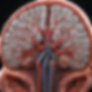
CT angiography (CTA) of the brain represents a crucial advancement in neuroimaging. It employs computed tomography to visualize blood vessels in the brain, providing insights into various cerebrovascular conditions. This technique significantly enhances diagnostic accuracy, making it an essential tool for clinicians. In this article, we will explore pertinent aspects of brain CT angiography, focusing on its methodologies, clinical applications, and associated safety protocols.
Research Overview
Summary of Key Findings
CTA provides detailed images of cerebral structures, allowing for the identification of conditions such as aneurysms, arteriovenous malformations, and occlusions. Studies show that CTA is highly sensitive and specific for detecting these vascular abnormalities compared to traditional methods like digital subtraction angiography.
Importance of the Research
The significance of CTA extends beyond diagnosis. It aids in treatment planning, monitoring disease progression, and evaluating the results of interventions. The growing prevalence of cerebrovascular diseases underscores the need for effective imaging techniques, making CTA a vital topic for ongoing research and clinical practice.
Methodology
Study Design
This article integrates findings from various studies to present a comprehensive overview of CTA practices. It includes both qualitative and quantitative research approaches that address the effectiveness and limitations of the technique in clinical settings.
Data Collection Techniques
Data for this overview were gathered through extensive literature reviews, examining peer-reviewed journals and contributions from neuroimaging conferences. This approach ensures that the information presented reflects the current state of knowledge in the field.
"CT angiography is revolutionizing the way we diagnose and manage cerebrovascular diseases."
The techniques employed in CTA involve the use of contrast agents, which enhance the visualization of blood vessels. The safety of these agents is critically assessed to mitigate potential risks to patients, ensuring a balance between diagnostic efficacy and patient safety.
As we delve deeper into the intricacies of brain CT angiography, it is essential to appreciate its role within the broader context of neuroimaging. Understanding the methodology and clinical relevance not only enhances diagnostic capability but also fosters advancements in patient care.
Prolusion to CT Angiography
CT angiography is an essential component in contemporary neuroimaging. It provides a clear window into the cerebral vasculature, allowing for enhanced diagnosis and treatment of various neurological issues. This section serves to lay the groundwork for understanding CT angiography, both its significance and its operational principles.
The role of CT angiography extends beyond simple imaging. It is instrumental in identifying abnormalities such as aneurysms, vascular malformations, and other cerebrovascular diseases. As such, its integration into the diagnostic pathway improves clinical decision-making and patient management. The non-invasive nature of the procedure also offers a significant advantage over traditional surgical methods, making it an attractive option for patients and healthcare providers.
Understanding CT Angiography
CT angiography utilizes advanced computed tomography technology to create detailed images of blood vessels in the brain. By employing a contrast agent, the technique enhances the visibility of these structures against surrounding tissues. This combination allows radiologists to identify irregularities that may indicate serious conditions.
The process begins when the patient receives an injection of a contrast material. This substance moves through the bloodstream, and as it does, the CT scanner captures multiple images from various angles. These images are then reconstructed to provide a three-dimensional representation of the vascular anatomy.
Additionally, CT angiography is notable for its rapid imaging capabilities. Unlike traditional angiography, which can be time-consuming and invasive, CT angiography can yield quick results, critical in emergency situations where timely intervention is vital.
History of CT Angiography Development
CT angiography has evolved significantly since its inception in the late 20th century. Initially, traditional angiography was the gold standard for visualizing blood vessels. However, the method involved invasive catheterization, which posed risks for patients. The development of CT technology in the 1970s laid the foundation for a less invasive approach.
The first applications of CT angiography began appearing in the early 1990s. Early systems and protocols faced limitations, including issues with image quality and dose of radiation. Over time, advancements in technology have dramatically improved the quality and speed of CT angiography.
Enhanced algorithms and sophisticated scanner models have now made it possible to visualize even minute vascular structures with remarkable clarity. The evolution of scanner capabilities continues to enhance the clinical utility of CT angiography in diagnosing and managing brain conditions.
Technical Principles of Brain CT Angiography
The technical principles underlying brain CT angiography are crucial for understanding its effectiveness and application in clinical settings. This section details the fundamental concepts that form the backbone of CT angiography technology, augmented by an exploration of the specific elements that contribute to its significance in neuroimaging. By grasping these principles, readers can appreciate the benefits this imaging modality provides in diagnosing and evaluating various neurological conditions.
Basic Concepts of CT Technology
CT angiography utilizes advanced computed tomography to deliver high-resolution images of blood vessels in the brain. Unlike traditional X-rays, which produce flat images, CT provides cross-sectional views, yielding a three-dimensional perspective of anatomical structures. This capability enhances the depth of image analysis, making it pivotal in identifying vascular conditions.
The working principle involves the use of rotating X-ray beams, which capture multiple images from various angles. A computer then reconstructs these images to create detailed cross-sectional views. This process, known as spiral or helical CT, allows for rapid acquisition of images and facilitates dynamic visualization of blood flow.
Effects of this technology include improved diagnostic accuracy and reduced time for image acquisition, leading to timely interventions when needed. Moreover, the versatility of CT angiography offers adaptability to various clinical needs, establishing its role as a cornerstone in neuroimaging.
Contrast Agents Used in CT Angiography
Contrast agents play an essential role in enhancing the visibility of blood vessels during CT angiography. These iodine-based substances are injected into the patient's bloodstream, allowing for the differentiation of vascular structures from surrounding tissues in the acquired images. The high atomic number of iodine facilitates this contrast, enhancing image clarity significantly.
However, the use of contrast agents necessitates careful consideration of patient-specific factors. Some individuals may experience allergic reactions or suffer from renal impairment, restricting the use of certain agents. Therefore, assessing patient history is vital before administering these substances.
Image Acquisition Techniques


The success of CT angiography hinges on efficient image acquisition techniques. Various modalities exist, each with distinct advantages and applications. Notably, single-phase and dual-phase CT angiography are common approaches.
- Single-Phase CT Angiography: This method captures images at a single point in time, typically during a peak enhancement of the vascular system. It is efficient for standard evaluations of arterial structures.
- Dual-Phase CT Angiography: This technique involves two consecutive imaging phases, which allows for evaluation of both the arterial and venous systems. It offers a more comprehensive view of cerebral vasculature.
Advances in technology have also introduced algorithms aimed at minimizing artifacts during image acquisition. For instance, techniques like iterative reconstruction enhance image quality while lowering radiation doses. Proper technique selection is crucial, depending on the clinical scenario at hand, ensuring the resulting images are both informative and diagnostically valuable.
Understanding these technical principles empowers healthcare professionals to harness the full potential of CT angiography in patient care.
Clinical Applications of Brain CT Angiography
CT angiography (CTA) of the brain is essential in the realm of modern neuroimaging. Its primary role is to provide precise visualization of cerebral blood vessels. The ability to assess these vessels critically aids in identifying various neurological conditions. This section delves into significant clinical applications of brain CTA, showcasing the tangible benefits it offers clinicians and patients alike.
Diagnosing Cerebrovascular Conditions
The diagnosis of cerebrovascular conditions is a foremost application of brain CT angiography. These conditions encompass a variety of disorders affecting blood flow to the brain, including stroke, arteriovenous malformations (AVMs), and carotid artery disease.
By utilizing CT angiography, medical professionals can gain insights into the vascular architecture of the brain. This imaging technique enables the rapid identification of blockages and abnormalities. For instance, in the case of acute ischemic stroke, time is critical; CTA can promptly visualize occluded arteries, facilitating timely therapeutic interventions.
Furthermore, CTA's ability to render high-resolution images aids in planning surgical or endovascular treatments.
- Convenience: It offers a quicker alternative compared to traditional imaging methods.
- Immediate results: Clinicians can assess the condition without extensive delays.
Evaluating Intracranial Aneurysms
Intracranial aneurysms represent localized dilations of blood vessels in the brain. These aneurysms can lead to significant complications if they rupture. CT angiography is particularly valuable in detecting and characterizing these vascular anomalies.
With its detailed imaging capabilities, CTA can showcase not only the presence of an aneurysm but also its size and morphology. Such information is critical in assessing the risk of rupture and determining management strategies.
In addition to the detection of existing aneurysms, CTA is instrumental in assessing the results following surgical or endovascular procedures. Surveillance imaging post-treatment ensures that recurrences or new aneurysms can be promptly identified.
Assessment of Vascular Malformations
Brain CT angiography is equally significant in evaluating vascular malformations, such as AVMs and cavernous malformations. These abnormalities can lead to serious outcomes, including hemorrhage and neurological deficits.
Through CTA, clinicians can visualize the intricate relationships between arteries and veins in malformations, which helps in understanding their dynamics. This imaging technique provides information on the size, location, and hemodynamics of vascular malformations.
It serves multiple roles:
- Initial diagnosis: Quickly confirms the presence of a malformation.
- Surgical planning: Offers crucial data needed for effective intervention.
- Monitoring: Keeps track of any changes in the vascular structure over time.
Advantages of Using CT Angiography
CT angiography (CTA) has transformed neuroimaging, offering several significant advantages that advance our ability to diagnose and manage cerebral vascular conditions. Understanding these benefits is essential for healthcare professionals, students, and researchers alike.
Rapid Imaging and Diagnosis
One of the primary advantages of CT angiography is its ability to provide rapid imaging and diagnosis. Unlike traditional angiography, which often requires an extensive setup and waiting time, CTA can acquire images within minutes. This immediacy plays a critical role in emergency situations where time is of the essence, such as during a suspected stroke. With quick access to high-quality images of the brain's vasculature, clinicians can promptly identify abnormalities such as occlusions or ruptured aneurysms.
This speed helps in making vital decisions about treatment pathways. For example, if a blood clot is detected in a patient with acute ischemic stroke, fast action can be taken, including the possible administration of thrombolytic therapy. Therefore, the efficiency of CT angiography significantly contributes to improved patient outcomes.
Non-Invasiveness of the Procedure
Another key advantage of CT angiography is its non-invasive nature. Patients do not require the use of catheters or other invasive devices as in conventional angiography, where access is gained through arterial puncture.
This non-invasiveness reduces the risks associated with the procedure, such as bleeding, infection, or vascular injury. Additionally, with no need for long recovery times, patients can often resume their normal activities quickly.
Furthermore, this advantage makes CTA a preferable option for patients who may be at higher risk due to existing medical conditions or those who are apprehensive about invasive techniques. In situations where repeated imaging is necessary, CTA provides a safer option.
Enhanced Visualization of Brain Vasculature
CT angiography also excels in providing enhanced visualization of brain vasculature. Using advanced imaging technology, CTA allows for detailed and rapid assessment of cerebral blood vessels. The high-resolution images generated enable radiologists to visualize complex vascular structures with remarkable clarity.
This level of detail can lead to improved identification of structural anomalies, including vascular malformations, atherosclerosis, and other conditions affecting cerebral blood flow. Clinicians can better understand the extent and nature of lesions, aiding in treatment planning.
"Through its unique imaging capabilities, CTA serves as a crucial tool in the assessment and management of cerebrovascular diseases."
Additionally, CTA's ability to produce 3D reconstructions of vascular systems allows for the evaluation of relationships between vessels and surrounding brain structures, which is invaluable during surgical planning or interventional radiology procedures.
In summary, the advantages of using CT angiography lie in its rapid imaging and diagnosis, non-invasive nature, and enhanced visualization of brain vasculature. Each of these elements not only supports clinical decision-making but also plays a critical role in ultimately improving patient care.
Limitations of CT Angiography
CT angiography (CTA) is a significant advancement in neuroimaging, but it is essential to explore its limitations. Understanding these constraints is imperative for clinicians to make informed decisions regarding patient diagnosis and management. Identifying the limitations allows healthcare professionals to weigh the benefits against potential drawbacks, ensuring optimal patient care.


Radiation Exposure Concerns
One prominent limitation of CT angiography is radiation exposure. While CTA provides valuable imaging information, the procedure involves the use of X-rays, which can pose risks, especially with repeated examinations. High doses of radiation can increase the likelihood of developing cancer over a person's lifetime. It's vital for practitioners to consider alternative imaging modalities, particularly in populations requiring frequent monitoring. Furthermore, dose-reduction techniques have been developed, but they may result in a compromise in image quality.
Few key points include:
- Patient education: Clinicians should discuss the risks with patients, allowing them to understand the benefit-risk ratio of undergoing CTA.
- Dose Optimization: Implementing protocols that minimize exposure without sacrificing image integrity is crucial.
- Regular Monitoring: Keeping track of radiation history helps to evaluate cumulative exposure over time.
"Understanding radiation exposure is critical in making judicious decisions in imaging practices."
Limitations in Specific Patient Populations
Certain populations may present additional challenges when it comes to the usefulness of CT angiography. For instance, patients with impaired renal function face risks associated with contrast agents used during the procedure. The iodinated contrast material can potentially worsen kidney function. In such cases, non-contrast imaging or alternative techniques like MRI angiography may be considered.
Moreover, factors like age and body habitus can affect imaging outcomes. Elderly patients or those with obesity may not only experience difficulty in maintaining the correct positioning for accurate imaging but may also yield suboptimal results due to artifacts.
Key considerations include:
- High-risk individuals: Evaluate renal function prior to the procedure.
- Positioning: Special attention may be required to achieve optimal scans for individuals with mobility limitations.
- Alternate Imaging Techniques: Keep different imaging modalities in mind to accommodate patient-specific conditions.
Potential Artifacts in Imaging Results
Artifacts in imaging are another notable limitation that can affect the interpretation of CT angiography results. Various factors can introduce artifacts during the scan, potentially masquerading as pathological findings. Movement artifacts, often due to inadequate patient cooperation or positioning, can lead to misinterpretations.
Moreover, metallic objects such as dental fillings or orthopedic implants may also produce distortions in the imaging output. These artifacts can obscure critical anatomical details, leading to complications in diagnoses. It is incumbent upon the radiologist to distinguish between genuine vascular abnormalities and imaging artifacts.
To mitigate these concerns, it is essential to:
- Educate patients about the importance of remaining still during the scanning process.
- Recognize potential artifacts associated with metallic devices.
- Use advanced software that may help reduce the impact of artifacts on final images.
In summary, while CT angiography remains an invaluable tool in diagnosing cerebrovascular conditions, understanding its limitations is crucial for successful application. Awareness of radiation exposure, challenges in specific patient populations, and potential imaging artifacts can guide clinicians in making more informed decisions.
Comparative Analysis with Other Imaging Modalities
The analysis of CT angiography in comparison to other imaging modalities holds significant relevance within this article. Understanding these variations can assist healthcare professionals in making informed decisions regarding which imaging technique to utilize based on clinical scenarios. Each imaging modality has its distinct advantages, limitations, and specific contexts where it excels.
CT Angiography vs. MRI Angiography
CT angiography (CTA) and MRI angiography (MRA) are far-too-often considered interchangeable, yet they exhibit crucial differences that impact their clinical use. CTA utilizes X-rays to produce images of blood vessels, with the added benefit of rapid data acquisition. This is especially important in acute settings, such as trauma, where timely diagnosis can significantly alter patient outcomes.
In contrast, MRA uses magnetic fields and radiofrequency pulses to create images without exposing the patient to ionizing radiation. While this may appear safer in certain contexts, MRI can have limitations such as longer scan times and reduced patient compatibility, particularly for those with implants or devices.
Both modalities excel in visualizing cerebral vasculature. However, CTA tends to provide superior spatial resolution and detailed bone assessment. This allows for a more accurate representation of vascular structures and potential obstructions.
In summary:
- CTA is faster and offers higher resolution for acute assessments.
- MRA avoids radiation, making it favorable for certain patients.
- Selection often depends on the specific clinical context and patient needs.
CT Angiography vs. Traditional Angiography
Traditional angiography, often referred to as digital subtraction angiography (DSA), remains the gold standard in certain vascular assessments. This procedure involves catheter insertion into the vascular system for direct contrast injection, followed by imaging. While this can yield highly detailed images and allow for therapeutic interventions, it is also invasive and carries inherent risks such as bleeding or infection.
CT angiography, on the other hand, is non-invasive, which provides a significant advantage in many situations. CTA can visualize vascular structures without catheterization. This can be particularly appealing when evaluating large populations or those at higher risk for complications. Moreover, CTA has become a common initial diagnostic tool due to its speed and ease of accessibility in emergency settings.
However, traditional angiography remains superior in assessing certain conditions, such as complex vascular anatomy or interventional radiology. In these cases, DSA allows for both diagnosis and immediate treatment.
Key differences include:
- Invasiveness:
- Risk Factors:
- Use Cases:
- CTA is non-invasive, while traditional angiography involves catheterization.
- CTA carries less risk and is generally safer for patients.
- Traditional angiography may be preferred for complex or interventional situations.
The choice between these options largely depends on the specific clinical scenario, the urgency of the imaging required, and patient considerations.
The comparative analysis of CT angiography with MRI angiography and traditional angiography demonstrates the necessity of context-specific decision-making in neuroimaging.
Patient Preparation for CT Angiography


Proper patient preparation for CT angiography is vital for both the accuracy of the imaging and the safety of the patient. This section discusses pre-procedure instructions and the assessment of allergies and medical history. It aims to elucidate why these aspects are essential components of the CT angiography process.
Pre-Procedure Instructions
Before undergoing CT angiography, patients must follow specific pre-procedure instructions to ensure optimal imaging results. First, it is crucial for patients to be informed about the need for fasting. Patients are often advised to refrain from eating or drinking for several hours before the procedure. This helps minimize the risk of nausea and ensures that the contrast agent administered later is more effective.
In addition to fasting, patients should be instructed to wear comfortable, loose-fitting clothing and to avoid metallic accessories. Items such as jewelry and belts can interfere with imaging quality and should be left at home or removed prior to the procedure.
Lastly, patients must also be educated on the importance of disclosing any medications currently being taken. Certain medications may affect the results of the scan or interact negatively with the contrast agent that is used. This can prevent complications post-procedure, contributing to a smoother workflow and better outcomes.
Assessment of Allergies and Medical History
Identifying allergies and reviewing medical history is a critical step in patient preparation for CT angiography. This assessment helps mitigate the risk of adverse reactions to contrast agents, which are commonly iodine-based.
- Allergy Screening: Clinicians should systematically inquire about any known allergies, especially to iodine, shellfish, or contrast media. A history of allergic reactions can necessitate alternative imaging options or premedication strategies to alleviate potential risks.
- Medical History: A comprehensive history includes discussing pre-existing medical conditions, such as renal insufficiency. Patients with poor kidney function face increased risks when exposed to contrast agents. Therefore, it may be necessary to conduct a kidney function test before proceeding with the scan.
These assessments ensure both the safety and efficacy of the CT angiography, allowing for tailored preparations specific to each patient’s needs.
"Thorough patient preparation not only enhances imaging quality but also safeguards the health of the individual undergoing the procedure."
Both pre-procedure instructions and an assessment of allergies and medical history are fundamental in optimizing the patient experience during CT angiography. They enhance the reliability of diagnostic results and reduce the risk of complications.
Safety and Risk Management
In the context of CT angiography, safety and risk management are crucial. As the technique involves the use of ionizing radiation and contrast agents, it is essential to consider both patient safety and the potential risks involved in the procedure. Effective management can significantly reduce the likelihood of adverse events, ensure high-quality imaging, and promote a positive experience for patients and healthcare providers alike.
Understanding Contrast Reactions
Contrast agents are commonly used in CT angiography to enhance the visualization of blood vessels. While these agents improve diagnostic accuracy, they do carry the risk of allergic reactions. The spectrum of reactions may range from mild symptoms like itching and rash to severe anaphylactic reactions, which can be life-threatening.
Healthcare professionals must be vigilant in assessing patient histories for known allergies to contrast media. Pre-procedure screening is essential. Patients with a history of allergies should be approached with caution. Additionally, protocols should be in place for immediate response to any reactions that occur during the imaging process. Providing patient education about potential reactions can also help to alleviate concerns and promote trust in the healthcare system. Understanding the patient's background can lead to better tailored approaches and safer outcomes.
Minimizing Radiation Risks
The use of CT technology inherently involves exposure to radiation. Therefore, mitigating radiation risks is paramount. The principle of ALARA, which stands for "As Low As Reasonably Achievable," serves as a guiding approach. This principle advocates for keeping radiation doses low while achieving high-quality images.
Several strategies exist to minimize radiation exposure:
- Optimizing Protocols: Utilizing appropriate scanning protocols tailored to specific clinical needs.
- Using Advanced Technology: Employing advanced CT machines that come equipped with dose-reduction features.
- Prioritizing Indications: Ensuring that CT angiography is performed only when necessary based on clinical indications.
By adhering to these practices, healthcare professionals can maintain diagnostic efficacy while prioritizing patient safety. Patient education about the importance of following pre-procedure instructions can also reduce unnecessary repeat scans, contributing to further reduction in radiation exposure.
In summary, safety and risk management are fundamental components of CT angiography practices. A comprehensive understanding of contrast reactions and radiation risks can significantly enhance the safety and effectiveness of this important diagnostic tool.
Emerging Technologies in CT Angiography
Emerging technologies in CT angiography represent a crucial advancement in neuroimaging. As the field of medical imaging evolves, these technologies aim to enhance visualization, improve accuracy, and reduce patient risk. Innovations in hardware and software have the potential to dramatically transform diagnostic capabilities. Understanding these advancements is essential for both clinicians and researchers, as they impact patient outcomes and the efficiency of medical practices.
Advancements in Imaging Techniques
Recent advancements in imaging techniques have led to more precise CT angiography. Techniques such as dual-energy imaging help to differentiate between various tissue types by using multiple energy levels. This can improve the contrast between blood vessels and surrounding tissues, making vascular structures clearer.
Another promising approach is the use of iterative reconstruction algorithms. These algorithms reduce image noise, allowing for clearer images at lower radiation doses. This helps address the concern of radiation exposure while maintaining high-quality imaging. Manufacturers like Siemens and GE Healthcare are at the forefront, continually integrating these technologies into their systems.
Additionally, the integration of artificial intelligence in image analysis holds great promise. AI can assist in identifying abnormalities in scans, thereby increasing diagnostic confidence and potentially reducing interpretation times. This kind of technology could significantly influence the workflow in busy imaging departments.
Future Directions in Neuroimaging
The future of neuroimaging, particularly in CT angiography, is expected to focus on several key areas. Increased collaboration between software developers and radiology departments will likely yield more tailored imaging solutions. Customization of imaging protocols for individual patients may become more common, optimizing the balance between image quality and dosage.
Moreover, the incorporation of real-time imaging during procedures is an area of active research. This could allow for immediate feedback and adjustments during surgical interventions, enhancing procedural efficacy.
The emergence of portable imaging units is another exciting frontier. These units could expand access to CT angiography in various settings, including remote or underserved areas. Such developments can play a pivotal role in early diagnosis and treatment of cerebrovascular conditions.
"The incorporation of AI in imaging not only enhances accuracy but also sets the stage for the future of personalized medicine in neuroimaging."
The End
CT angiography of the brain plays a crucial role in modern neuroimaging. Its ability to provide detailed images of cerebral vessels aids in diagnosing many neurological conditions. This imaging technique stands out for its speed and non-invasiveness, which can lead to timely treatments for patients.
Several key points highlight the significance of this article's conclusion:
- Summary of Core Benefits: The advantages of CT angiography are manifold. One such benefit is the rapid imaging capability, which allows clinicians to make swift decisions in urgent situations.
- Clinical Relevance: Understanding the applications of this imaging is vital for advancing patient care. It is important for healthcare professionals to stay informed about how CT angiography enhances diagnostic accuracy for cerebrovascular diseases.
- Continued Research: As technology advances, staying updated with the latest developments in CT angiography can significantly impact diagnostic practices and outcomes. This article serves as a roadmap for future studies on emerging techniques and improvements.
Furthermore, while CT angiography offers significant benefits, it is essential to acknowledge its limitations, including radiation exposure and potential artifacts in imaging. These considerations must be factored into clinical decisions for patient safety.
"Knowledge equips us to navigate the complexities of diagnostic science and enhance patient outcomes."
By synthesizing the information presented throughout this article, the reader gains a comprehensive perspective on the importance of CT angiography in contemporary neuroscience.


